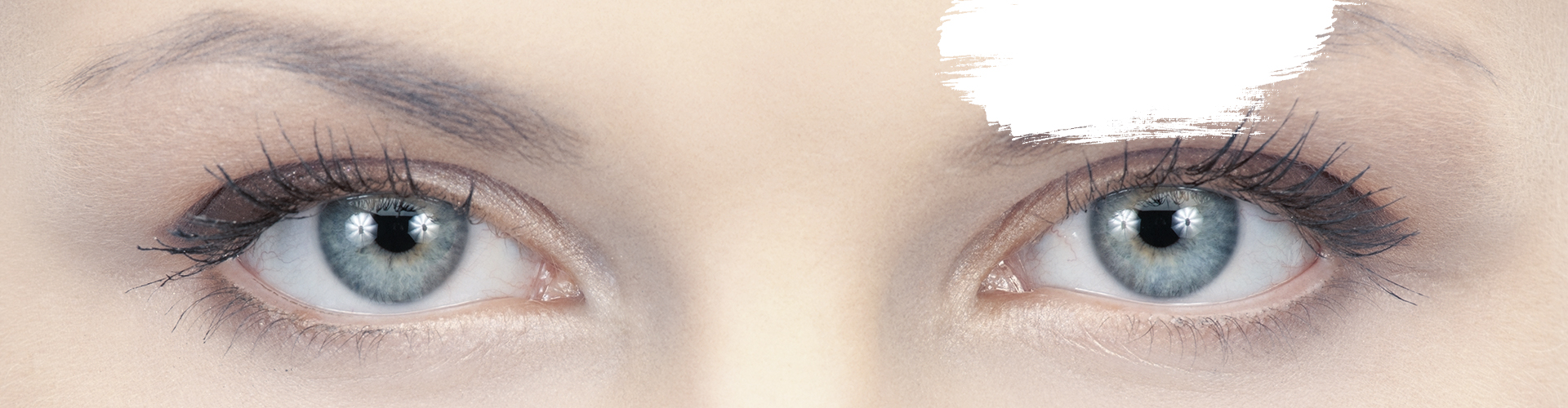
Take a Closer Look at Your Eyes
Your eyes are a complex network of intricate systems, all of which work together to bring you your gift of sight.
To better understand the issues your eyes might face, it’s sometimes best to look at these systems in detail. By helping you understand how your eyes work, we hope to encourage you to take care of your eyes and have regular eye exams.
But the best way to understand your unique set of eyes and their needs is to get them examined by our doctors. Book an eye exam with our team today!
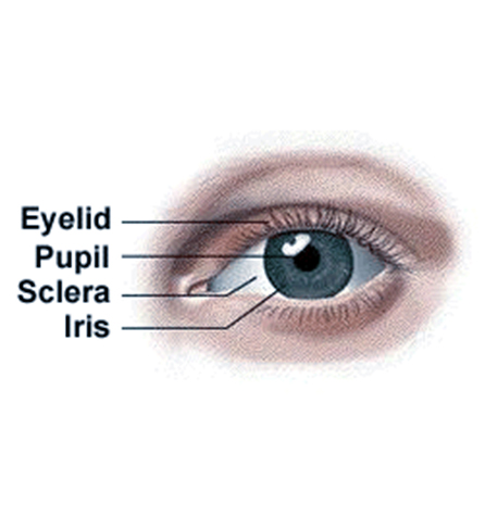
Taking a Look at Your Eye
There is a lot to discover when it comes to your eyes. Each part has a very important role to play ranging from comfort, to visual clarity, all the way to helping you see colors. While there are many different working parts your eye’s use, some of the most important include:
The Conjunctiva
The conjunctiva is a clear membrane that covers parts of your eye’s front surface as well as the inner surface of your eyelids. It’s responsible for keeping your eyes lubricated and free from debris, allowing your eyelids to glide comfortably across your eye’s surface.
However, issues like conjunctivitis can cause irritation, eye pain, or even mucusy discharge.
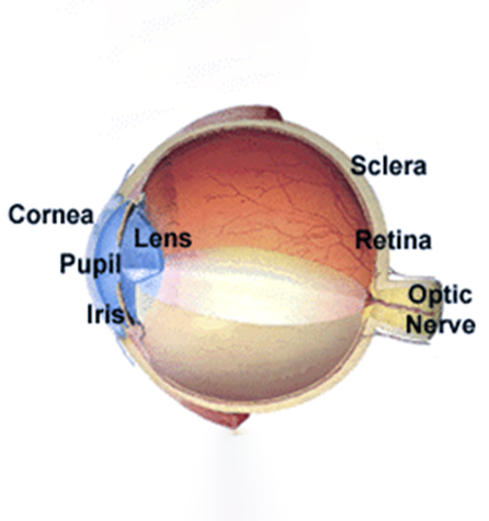
The Sclera
The sclera is more widely known as the whites of your eyes. The sclera makes up 80% of your eye’s total surface area and extends from your cornea all the way to your optic nerve at the back of the eye.
Alongside your intraocular pressure, the sclera helps maintain your eyes’ shape. The sclera also attaches to exterior muscles that help control your eye movement.
The Cornea
If you have ever heard the term “the eyes are the window to the soul,” the cornea would be the window.
The cornea is located right in front of your iris and pupil and is largely responsible for focusing the light that enters your eye. The cornea features several different layers, the most notable being:
- The epithelium: The epithelium is your cornea’s outermost layer. This layer can help protect your eyes from more superficial injuries and is a vital surface for your tear film to spread over.
- The stroma: The stroma is the middle layer of your cornea and its largest, making up about 90% of your cornea’s thickness. Your stroma is what ophthalmologists reshape during laser eye surgery to help correct your vision.
- The endothelium: The endothelium is the innermost layer of your cornea. This layer is vital for maintaining the fluids in your cornea and keeping your cornea clear.
The Pupil & Iris
Up next, we have the pupil and iris. The pupil is the black circle in the cornea’s middle, while the iris is the colored portion surrounding it. Your iris controls the size of your pupil, which in turn determines how much light enters your eyes at any given moment.
For example, if you’re in a bright environment, your iris will contract the size of your pupil to limit the light reaching your retina, and vice versa for when you’re in darker environments.
The Lens
Right behind your pupil is your lens, a clear structure responsible for providing additional focusing powers.
Your lens is responsible for bending and focusing light onto your retina. It does this by stretching or shrinking a number of ciliary muscles whenever you look at an object.
Some of the most common issues your lens can develop include presbyopia and cataracts, both of which can occur with age.
The Retina
The retina is the large area at the back of your eye that receives the light that passes through the lens and cornea.
The retina features a layer of cells called photoreceptors responsible for taking light and turning it into chemical and nervous signals that pass through your optic nerve to your brain. This process provides the vision you’re using right now.
However, damage to these photoreceptors, and your retina in general, is permanent. Common issues that can damage your retina include diabetic retinopathy and UV overexposure.
The Macula
The macula is the most light-sensitive part of your retina, and it’s responsible for providing you with clear central vision and color vision.
Doctors can observe your macula during a comprehensive eye exam, which is essential for detecting common issues like age-related macular degeneration and diabetic macular edema.
The Optic Nerve
Your optic nerve is responsible for carrying the information your retina collects to your brain. Your optic nerve connects to the back of your eye. Doctors can observe the connection between your retina and optic nerve by looking at your optic disc, a circular part of your retina that signifies the beginning of your optic nerve.
One of the most common issues the optic nerve might experience is glaucoma, a group of eye diseases that can damage your optic nerve over time. Doctors can diagnose these issues during an eye exam.

Learn More About Your Eyes Today
The very first step to managing your eye health is to learn what your eyes need. We can help discover your eye’s unique intricacies during an eye exam while also taking proactive measures to maintain and preserve your eye health and vision.
Visit Us Today
Where to Find Us?
You can find us on Warner Avenue just over a mile from the beach. We have plenty of parking out front!
Our Address
- 5092 Warner Avenue
- Huntington Beach, CA 92649
Contact Information
- Phone: (714) 846-2897
- Email: huntingtonbeach@totalvisionllc.com
Hours of Operation
- Monday: 9:00 AM – 5:00 PM
- Tuesday: 9:00 AM – 5:00 PM
- Wednesday: 9:00 AM – 5:00 PM
- Thursday: 9:00 AM – 5:00 PM
- Friday: 9:00 AM – 5:00 PM
- Saturday: Closed
- Sunday: Closed
Our Eye Care Services

Advanced Contact Lens Care

Comprehensive Eye Exams

Keratoconus

Children’s Eye Exams
Our Brands


Our Google Reviews
Be the First to Know,
Be the First to Win.
From eye health insights to exclusive giveaways, your feed just got a lot clearer.

-
Total Vision - Huntington Beach
- 5092 Warner Avenue
- Huntington Beach, CA 92649
-
P: (714) 846-2897
huntingtonbeach@totalvisionllc.com